
Can an MRI Detect a Pinched Nerve?
Yes, an MRI can help identify a pinched nerve, particularly when a nerve is compressed by a herniated disc, spinal stenosis, bone changes, or another structural problem. MRI provides detailed images of the spine and surrounding soft tissues, helping doctors identify possible nerve-root compression.
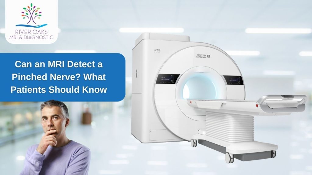
Quick Answer
An MRI can show structural causes of nerve compression, such as a herniated disc, spinal stenosis, or narrowing around a nerve. However, MRI does not directly measure how well a nerve is functioning. When nerve function needs to be evaluated, a doctor may also recommend tests such as EMG or nerve conduction studies.
A pinched nerve can cause symptoms such as pain, numbness, tingling, burning sensations, or weakness. MRI can help identify whether a structural abnormality may be contributing to those symptoms and where the compression may be occurring.
For patients with suspected nerve compression, the appropriate imaging depends on the location of symptoms and the clinical question. A cervical spine MRI may be used for symptoms involving the neck and arm, while a lumbar spine MRI may be considered for lower-back and leg symptoms.
What Is a Pinched Nerve?
A pinched nerve occurs when surrounding tissue puts pressure on a nerve. In the spine, this may involve pressure on a spinal nerve root and can lead to symptoms such as pain, numbness, tingling, burning sensations, or weakness.
When nerve compression occurs in the spine, doctors may refer to it as radiculopathy. The symptoms can vary depending on which nerve root is affected and whether the compression occurs in the neck or lower back.
What Happens When a Nerve Gets Compressed?
When a nerve is compressed or irritated, it can interfere with the signals traveling between the nerves and the rest of the body. This may cause pain that travels along the affected nerve, along with numbness, tingling, or weakness.
For example, compression of a nerve root in the neck may cause symptoms extending into the shoulder, arm, or hand. Compression in the lower back can cause pain or other symptoms that travel into the buttock or leg, including symptoms commonly associated with sciatica.
MRI can be particularly useful when the suspected problem involves spinal nerve compression, because it provides detailed images of the spine, discs, and surrounding soft tissues.
What Causes a Pinched Nerve?
Several structural changes can put pressure on a spinal nerve or nerve root. Common causes include:
- Herniated disc: Part of a spinal disc can extend beyond its normal position and press against a nearby nerve.
- Bulging disc: A disc can bulge outward as it changes over time. A bulging disc does not always cause symptoms, but it can contribute to nerve irritation or compression depending on its location.
- Spinal stenosis: Narrowing within the spinal canal or areas where nerves exit can reduce the available space around the nerves. MRI can help show changes involving the discs and other spinal structures.
- Bone spurs: These bony growths can develop along the spine and sometimes narrow the spaces where spinal nerves travel.
- Narrowing around nerve roots: Changes in the spine can reduce the space around a nerve root and contribute to compression.
- Other structural changes: Degenerative changes, injuries, or other abnormalities may also affect the space around a nerve.
Because several conditions can produce similar symptoms, identifying the possible source of MRI nerve compression or MRI nerve root compression requires more than symptoms alone. A healthcare professional may use a physical examination along with imaging or other tests to determine what is causing the problem.
How Does an MRI Detect a Pinched Nerve?
An MRI does not “see” a pinched nerve in the same way a camera sees an object. Instead, it creates detailed images of the spine, discs, spinal canal, and surrounding soft tissues. These images can help doctors identify structural changes that may be pressing on or irritating a nerve root.
MRI is commonly used when a healthcare professional suspects nerve-root compression, particularly when symptoms suggest a problem involving the spine.
What Does a Pinched Nerve Look Like on an MRI?
A pinched nerve may appear on an MRI as a nerve root being crowded, displaced, or compressed by a nearby structure. The images can show abnormalities such as a herniated disc, spinal stenosis, bone spurs, or other changes that reduce the space available for a nerve.
For example, a herniated disc can extend into the space occupied by a spinal nerve root. MRI can show the disc abnormality and its relationship to nearby neural structures, helping the interpreting radiologist assess whether the finding could explain the patient’s symptoms.
However, an MRI finding does not automatically mean that it is the source of a patient’s pain. Some spinal changes can be present without causing symptoms, so MRI findings are considered alongside the patient’s symptoms and physical examination.
What Does Nerve Compression Look Like on an MRI?
MRI nerve compression can appear as reduced space around a nerve root, direct contact between a disc or other structure and the nerve, or narrowing of the passage through which a nerve exits the spine.
The exact appearance depends on where the compression occurs. In the neck, for example, changes in the cervical spine may affect nerve roots traveling toward the shoulder, arm, or hand. In the lower back, compression of a lumbar nerve root can contribute to radiating pain, numbness, tingling, or weakness in the leg.
Because MRI provides detailed soft-tissue images, it can help evaluate both the structures causing the narrowing and the location of suspected MRI nerve root compression.
What MRI Can Show: MRI provides detailed anatomical images that can help identify structural causes of nerve compression, including herniated discs, narrowing around nerve roots, and other spinal changes.
Can MRI Show the Cause of Nerve Compression?
Yes. One of the main advantages of MRI is that it can help identify the structural cause of nerve compression, rather than simply showing that a patient has pain.
Depending on the clinical situation, MRI may help identify:
- Herniated or bulging discs
- Spinal stenosis
- Bone spurs
- Narrowing around spinal nerve roots
- Degenerative changes in the spine
- Other structural abnormalities affecting the spinal canal or nerve pathways
This makes MRI useful when a doctor needs to investigate whether a structural problem could be contributing to symptoms such as radiating pain, numbness, tingling, or weakness. Mayo Clinic lists MRI among the tests that may be used when nerve-root compression is suspected.
For patients who need diagnostic imaging, RiverOaks MRI & Diagnostic provides MRI services in Sugar Land, including imaging for conditions involving the spine and surrounding soft tissues.
Can MRI Detect a Pinched Nerve in the Neck?
Yes, an MRI can help identify a pinched nerve in the neck by showing changes in the cervical spine that may compress or irritate a nerve root. A cervical spine MRI provides detailed images of the discs, spinal canal, neural foramina, and surrounding soft tissues, helping doctors evaluate possible cervical nerve compression.
Cervical Spine MRI for Nerve Compression
A cervical spine MRI focuses on the neck portion of the spine. It can help show structural problems that may affect the nerve roots, including herniated or bulging discs, spinal narrowing, bone spurs, and other degenerative changes.
When a nerve root is compressed in the neck, the condition is commonly referred to as cervical radiculopathy. The symptoms can extend beyond the neck because the affected nerve roots supply areas of the shoulder, arm, and hand.
For chronic neck pain accompanied by radiculopathy, the American College of Radiology states that an MRI of the cervical spine without contrast is usually an appropriate initial imaging study and is particularly useful for assessing suspected nerve-root impingement.
Symptoms That May Lead to a Neck MRI
Not every case of neck pain requires an MRI. However, imaging may be considered when symptoms suggest possible nerve compression or when symptoms persist or require further evaluation.
Common symptoms can include:
- Neck pain that may extend into the shoulder
- Arm pain that travels away from the neck
- Tingling or pins-and-needles sensations
- Numbness in the arm or hand
- Weakness in the arm or hand
- Pain radiating into the hand or fingers
These symptoms can occur when a cervical nerve root is compressed or irritated, although they can also have other causes. A physical examination and the patient’s overall symptoms help determine whether a cervical spine MRI for a pinched nerve is appropriate.
Quick Answer: If neck pain is accompanied by arm pain, numbness, tingling, or weakness, a cervical spine MRI may help identify structural causes of nerve compression, such as a herniated disc or narrowing around a nerve root.
If imaging is recommended, the MRI findings should be interpreted together with the patient’s symptoms and examination. An abnormality on a scan does not necessarily mean it is the source of every symptom.
Can MRI Detect a Pinched Nerve in the Lower Back?
Yes, an MRI can help identify a pinched nerve in the lower back by showing structural changes that may compress a lumbar nerve root. A lumbar spine MRI provides detailed images of the discs, spinal canal, nerve-root pathways, and surrounding soft tissues, helping clinicians evaluate possible causes of radiating pain, numbness, tingling, or weakness.
Lumbar MRI for Nerve Compression
A lumbar spine MRI focuses on the lower portion of the spine, where nerve-root compression can cause symptoms in the lower back, buttocks, legs, or feet.
Common structural causes include herniated or bulging discs, spinal narrowing, bone changes, and other abnormalities that reduce the space around a nerve root. When a spinal nerve root is compressed in the lower back, it is commonly described as lumbar radiculopathy.
MRI can show these anatomical changes and their relationship to nearby nerve structures. However, an abnormal finding does not necessarily mean it is causing a person’s symptoms. MRI results need to be interpreted alongside the patient’s symptoms and physical examination.
MRI for Sciatica and Leg Symptoms
Sciatica describes pain that travels along the pathway of the sciatic nerve, typically from the lower back or buttock into the leg. A herniated disc or other changes affecting lumbar nerve roots can cause sciatica. Symptoms may include radiating leg pain, numbness, tingling, or muscle weakness.
An MRI for sciatica can help identify structural problems such as a herniated disc or other changes that may be affecting a lumbar nerve root. Mayo Clinic notes that MRI can produce detailed images of soft tissues and show herniated discs and pinched nerves.
However, not everyone with lower-back pain or sciatica needs an MRI immediately. The American College of Radiology notes that imaging is generally not appropriate as the initial test for uncomplicated acute low-back pain or radiculopathy without red flags. MRI becomes more appropriate in certain situations, such as persistent or progressive symptoms when a patient may be a candidate for surgery or another intervention.
Quick Answer: A lumbar MRI can help show whether a herniated disc, spinal narrowing, or another structural change may be compressing a nerve root and contributing to sciatica or other leg symptoms.
What Symptoms May Mean You Need an MRI for a Pinched Nerve?
Certain symptoms can suggest that a nerve is irritated or compressed, particularly when they follow a consistent path from the neck or back into an arm or leg. An MRI may be considered when a healthcare professional suspects that a structural problem, such as a herniated disc or spinal narrowing, could be affecting a nerve root.
Not everyone with these symptoms needs an MRI. The decision depends on the symptoms, physical examination, duration and severity of the problem, and the suspected underlying cause.
Numbness and Tingling
Numbness or tingling can occur when a nerve is compressed or irritated. You may notice reduced sensation, pins-and-needles, or an unusual feeling in an arm, hand, leg, or foot.
An MRI for numbness and tingling may help evaluate whether a structural problem in the spine could be affecting a nerve root. However, numbness and tingling can have many causes, so imaging is not automatically required for these symptoms.
Burning or Shooting Pain
A compressed nerve can cause burning, sharp, shooting, or electric-like pain. These sensations may follow the path of the affected nerve rather than staying in one location.
For persistent burning nerve pain or shooting pain, a healthcare professional may evaluate whether the symptoms could be related to nerve compression. When a spinal cause is suspected, MRI can help identify structural abnormalities that may be contributing to the symptoms.
Pain That Travels Into an Arm or Leg
Pain that starts in the neck or lower back and travels into an arm or leg can occur when a spinal nerve root is irritated or compressed.
For example, cervical nerve-root problems can cause symptoms extending into the shoulder, arm, or hand. Lumbar nerve-root compression can cause pain that travels from the lower back or buttock into the leg, as commonly seen with sciatica.
An MRI may help determine whether a disc problem, spinal narrowing, bone changes, or another structural abnormality could be affecting the nerve.
Muscle Weakness
Muscle weakness in an arm, hand, leg, or foot can occur when a nerve is affected. For example, you may notice difficulty gripping objects, lifting the foot, or performing movements that were previously easy.
Weakness deserves medical evaluation because it can have several possible causes. If a clinician suspects nerve-root compression, an MRI may help look for a structural cause. Other tests, including EMG or nerve conduction studies, may also be considered when information about nerve function is needed.
Quick Answer: Numbness, tingling, burning or shooting pain, radiating arm or leg pain, and muscle weakness can occur with nerve compression. When a spinal cause is suspected, an MRI may help identify structural changes that could be affecting the nerve.
Can an MRI Detect Nerve Damage?
MRI can show structural abnormalities associated with nerve problems, but it does not directly measure how well a nerve is functioning. For example, an MRI may identify a herniated disc, spinal narrowing, tumor, injury, or another structural change that affects a nerve. When the goal is to evaluate electrical nerve function or determine whether a nerve is damaged, additional testing such as EMG or nerve conduction studies may be used.
This distinction matters because “nerve compression” and “nerve damage” are not always the same thing. A compressed nerve may cause symptoms without permanent damage, while prolonged or significant compression can sometimes lead to nerve injury.
MRI vs. Tests That Assess Nerve Function
MRI and EMG answer different diagnostic questions.
MRI shows anatomy. It produces detailed images that can help identify structural causes of nerve compression, such as a herniated disc, spinal stenosis, or other changes affecting a nerve root. Mayo Clinic specifically lists MRI as a test that may be used when nerve-root compression is suspected.
EMG and nerve conduction studies assess function. EMG records electrical activity in muscles, while nerve conduction studies measure how electrical signals travel through nerves. These tests can provide information about nerve dysfunction or damage that an anatomical scan alone cannot establish.
So, when comparing MRI vs. EMG for a pinched nerve, neither test answers exactly the same question:
- MRI: What structural problem may be compressing or affecting the nerve?
- EMG/NCS: Is there evidence of abnormal nerve or muscle function?
Depending on the symptoms and suspected cause, a healthcare professional may use one test or both.
Can You Have a Pinched Nerve With a Normal MRI?
Yes, it is possible to have symptoms consistent with nerve irritation or compression even when an MRI does not show a clear structural cause. MRI findings also need to be interpreted in the context of the patient’s symptoms and physical examination.
An MRI may show a structural abnormality that is not actually responsible for a person’s symptoms. Conversely, symptoms can have causes that are not clearly demonstrated on a routine MRI. This is why doctors may combine imaging with neurological examination and, when appropriate, EMG or nerve conduction testing.
Quick Answer: MRI can help identify structural causes of nerve compression, but it does not directly measure nerve function. EMG and nerve conduction studies can provide additional information about whether nerves and muscles are functioning normally.
This means that if you are searching “can MRI detect nerve damage?” or “can MRI confirm a pinched nerve?”, the answer depends on what is causing the symptoms and what information the healthcare professional needs. MRI is particularly useful for looking for structural nerve compression, while nerve-function tests can provide complementary information.
MRI vs. EMG for a Pinched Nerve
MRI and EMG are different types of tests. MRI looks at anatomy and can help identify structural causes of nerve compression, while EMG evaluates electrical activity and provides information about nerve and muscle function. A doctor may recommend one or both depending on the symptoms, physical examination, and suspected cause. (mayoclinic.org)
| Test | What It Shows |
|---|---|
| MRI | Structural changes and possible nerve compression |
| EMG | Electrical activity and nerve/muscle function |
| X-ray | Bone structure and alignment |
| CT | Detailed cross-sectional anatomy |
When Is an MRI Used?
An MRI may be useful when a healthcare professional wants to investigate whether a structural problem is affecting a nerve. It can provide detailed images of discs, the spinal canal, nerve-root pathways, and surrounding soft tissues.
For example, an MRI may help identify a herniated disc, spinal stenosis, bone changes, or other abnormalities that could contribute to nerve compression.
When Is an EMG Used?
An EMG can provide information about electrical activity in muscles and help evaluate whether there is abnormal nerve or muscle function. Nerve conduction studies are often performed alongside EMG to assess how electrical signals travel through nerves. (mayoclinic.org)
This can be useful when symptoms suggest a nerve problem but the clinician needs more information about how the nerve is functioning.
MRI or EMG for Nerve Damage?
The answer depends on the diagnostic question.
If the concern is what structure may be compressing a nerve, MRI can provide valuable anatomical information. If the concern is whether the nerve is functioning abnormally, EMG and nerve conduction studies can provide complementary information.
In some situations, both may be recommended because they provide different types of information.
What About X-Ray and CT?
X-rays can show bones and alignment but provide less detail of nerves, discs, and other soft tissues than MRI. CT produces detailed cross-sectional images and can provide useful information about bone and other anatomical structures.
CT scans can provide detailed images of bones and other structures, while MRI is generally more useful for evaluating soft tissues, discs, the spinal cord, and structures around nerves. The right test depends on the symptoms and what your doctor is trying to identify. For a broader comparison, see our guide to MRI vs. CT scan.
The appropriate test depends on factors such as the patient’s symptoms, medical history, physical examination, suspected cause, and the specific question the clinician needs to answer. There is no single imaging or diagnostic test that is appropriate for every person with a suspected pinched nerve.
Quick Answer: MRI evaluates structural changes that may compress a nerve, while EMG evaluates nerve and muscle function. X-ray primarily shows bone structure, and CT provides detailed cross-sectional anatomy. Your healthcare professional can determine which test or combination of tests is appropriate for your symptoms.
Is MRI the Right Test for a Pinched Nerve?
MRI can be an important test when a doctor suspects that a pinched nerve is caused by a structural problem such as a herniated disc, spinal stenosis, or another change affecting a nerve root. However, MRI is not automatically the right test for every person with nerve pain, numbness, or tingling. The choice depends on your symptoms, physical examination, suspected cause, and how long the symptoms have been present.
When MRI May Be Useful
MRI may be useful when a doctor needs detailed images of the spine, discs, nerves, and surrounding soft tissues. It can help identify structural causes of nerve compression, including herniated discs and narrowing around nerve roots.
For example, MRI may be considered when symptoms such as radiating arm or leg pain, numbness, tingling, or weakness suggest possible nerve-root compression. In some cases, MRI is also used when symptoms persist or progress and additional imaging is needed to guide further treatment.
When Another Test May Be Needed
MRI shows anatomy, but it does not directly measure how well a nerve is sending electrical signals. If a doctor needs to evaluate nerve or muscle function, an EMG (electromyography) and nerve conduction study may provide additional information.
Other imaging tests can also have a role. X-rays can show bone alignment and certain structural changes, while CT can provide detailed images of bone and may be useful in specific situations. The appropriate test depends on what your healthcare provider is trying to determine.
Why Your Doctor Chooses a Specific Imaging Test
There is no single imaging test that is appropriate for every suspected pinched nerve. Your doctor may consider:
- Where your symptoms are located
- Whether pain, numbness, tingling, or weakness is present
- How long symptoms have lasted
- Results of your physical and neurological examination
- Whether there has been an injury or trauma
- Whether there are signs that require more urgent evaluation
- Whether the concern is structural nerve compression or nerve function
For example, MRI may be useful when the concern is nerve-root compression from a spinal problem, while EMG and nerve conduction studies can provide information about nerve and muscle function. Imaging findings also need to be interpreted alongside your symptoms and clinical examination rather than considered on their own.
Quick Answer: MRI can be an appropriate test for a suspected pinched nerve when a doctor needs to look for structural nerve compression. However, depending on your symptoms and the suspected cause, your doctor may recommend an EMG, X-ray, CT scan, or another diagnostic test instead.
What Happens During an MRI for a Pinched Nerve?
A spine MRI is a noninvasive imaging test. You will lie on a padded table that moves into the MRI scanner while detailed images of the spine and surrounding tissues are taken. The scan itself is generally painless, although you need to remain still while the images are captured.
Does a Spine MRI Hurt?
No. MRI does not normally cause pain. You may hear loud tapping or thumping sounds during the scan, and some people may feel uncomfortable staying still or experience claustrophobia.
If you want to understand what to do before your appointment, see our MRI preparation guide for a more detailed checklist.
How Long Does a Pinched-Nerve MRI Take?
A spine MRI typically takes about 30 to 60 minutes, depending on the area being scanned and the imaging protocol. If contrast is required, additional images may add time to the examination.
Do You Need Contrast?
Not always. Many spine MRI examinations can be performed without contrast. Your doctor and radiology team will determine whether contrast is appropriate based on your symptoms, medical history, and the reason for the examination.
MRI Safety Screening
Before an MRI, the technologist will ask about metal implants, medical devices, previous surgeries, and other safety considerations. You may also need to remove jewelry, watches, phones, and other metal objects before entering the MRI room. Tell the technologist about any implanted device or metal in your body before the scan.
For complete preparation instructions, patients can review RiverOaks MRI & Diagnostic’s MRI preparation guide before their appointment.
Open MRI or 3T MRI for a Pinched Nerve?
If your doctor recommends an MRI for a suspected pinched nerve, the type of MRI may depend on the body area being examined, the symptoms being evaluated, and the imaging protocol your doctor orders. Open MRI and 3T MRI offer different features, but one is not automatically the right choice for every pinched nerve.
Open MRI in Sugar Land, TX
An Open MRI uses a more open design that can provide a more comfortable experience for patients who feel anxious or uncomfortable in a traditional enclosed scanner. RiverOaks MRI & Diagnostic offers Open MRI in Sugar Land using the Hitachi AIRIS Elite system. The facility describes its Open MRI as suitable for imaging areas including the spine, joints, and soft tissues.
For patients searching for Open MRI in Sugar Land, TX or an Open MRI near me in Sugar Land, TX, an open design may be worth discussing with the imaging team, particularly if claustrophobia or scanner comfort is a concern.
3T MRI in Sugar Land, TX
A 3T MRI uses a stronger magnetic field than many conventional MRI systems and can produce detailed, high-resolution images. RiverOaks MRI & Diagnostic describes its 3T MRI as being used for imaging the brain, spine, joints, and soft tissues.
A 3T MRI may be considered when detailed imaging is needed, but higher field strength does not automatically mean it is the right choice for every pinched nerve. The appropriate MRI system and protocol depend on the body part being examined and the clinical question your doctor is trying to answer.
For patients looking for 3T MRI in Sugar Land, TX, 3 Tesla MRI in Sugar Land, TX, or an MRI center in Sugar Land, TX, the imaging team can help determine which available MRI option matches the examination ordered by your healthcare provider.
Internal link: Link 3T MRI to the RiverOaks 3T MRI service page.
Which MRI Is Right for a Pinched Nerve?
The choice between Open MRI and 3T MRI should be based on the examination needed, the area being scanned, and patient-specific considerations. Your doctor or imaging team can determine whether a particular MRI system and protocol are appropriate for evaluating suspected nerve compression.
MRI for a Pinched Nerve in Sugar Land, TX
If you have symptoms that may be related to a pinched nerve and your healthcare provider has recommended MRI imaging, choosing a convenient diagnostic imaging center can make the process easier. RiverOaks MRI & Diagnostic provides MRI and other diagnostic imaging services in Sugar Land, Texas.
RiverOaks MRI & Diagnostic in Sugar Land
RiverOaks MRI & Diagnostic is located at:
2121 Williams Trace Blvd, Suite 100
Sugar Land, TX 77478
Phone: 281-453-2055
The center is open Monday through Friday and provides diagnostic imaging services for patients in Sugar Land and surrounding communities.
If your doctor has recommended an MRI to evaluate possible nerve compression, the imaging team can provide information about the available MRI options and help with scheduling.
MRI, Open MRI and 3T MRI Services
RiverOaks offers several diagnostic imaging options, including MRI, Open MRI, 3T MRI, CT, X-ray, and ultrasound, along with pain management services.
For suspected nerve compression, the appropriate MRI system and imaging protocol depend on the body area being examined and the clinical question. RiverOaks provides:
- MRI Services for detailed diagnostic imaging
- Open MRI for patients who may prefer a more open scanning environment
- 3T MRI for high-resolution imaging, including imaging of the spine and other areas
Learn more about MRI Services, Open MRI, and 3T MRI to understand the available imaging options.
If you are looking for an MRI center in Sugar Land, TX for suspected nerve compression, RiverOaks MRI & Diagnostic is located conveniently at Williams Trace Blvd in Sugar Land.
Why Choose RiverOaks MRI & Diagnostic for MRI Imaging?
Choosing an imaging center involves more than finding a convenient appointment. The technology available, the type of MRI examination, patient comfort, and quality standards can all be important considerations.
RiverOaks MRI & Diagnostic combines advanced MRI technology, Open MRI options, 3T MRI imaging and ACR-accredited MRI services in Sugar Land, Texas.
Advanced MRI Technology
RiverOaks offers multiple MRI options designed for different imaging needs. Its MRI services include 1.5T MRI and Open MRI, while the center also offers 3T MRI for high-resolution imaging. The appropriate system depends on the body area being examined and the clinical question.
Open MRI Options
For patients who are uncomfortable in a traditional enclosed scanner, an Open MRI can provide a more open scanning environment. RiverOaks uses the Hitachi AIRIS Elite Open MRI system and offers Open MRI imaging in Sugar Land.
3T MRI Imaging
RiverOaks also provides 3T MRI, which uses a higher magnetic field strength to produce detailed images. The center describes its 3T MRI as suitable for imaging areas including the brain, spine, joints, and soft tissues.
For a suspected pinched nerve, the appropriate MRI system and protocol should be determined according to the examination being ordered rather than simply choosing the highest field strength.
Patient-Focused Imaging Experience
MRI requires patients to remain still during image acquisition, so comfort and communication can be important parts of the imaging experience. RiverOaks describes its MRI services as patient-centered and provides an Open MRI option for patients who may be uncomfortable with a more enclosed scanner.
ACR Accreditation
RiverOaks reports that its Open and Closed MRI machines have earned American College of Radiology (ACR) accreditation following a review process. ACR accreditation evaluates areas such as equipment quality, staff qualifications, image quality, and safety practices.
For patients researching an MRI provider, accreditation can provide useful information about the facility’s commitment to established imaging quality and safety standards.
A Factual Choice for MRI Imaging in Sugar Land
If your doctor has recommended an MRI for a suspected pinched nerve, RiverOaks offers multiple MRI options in Sugar Land, including Open MRI and 3T MRI. Your healthcare provider and imaging team can determine which examination and MRI protocol are appropriate for your symptoms and clinical needs.
Frequently Asked Questions About MRI and Pinched Nerves
Can an MRI Detect a Pinched Nerve?
Yes, an MRI can help detect a pinched nerve by showing structural problems that may compress a nerve, such as a herniated disc, spinal stenosis, or other changes around the spine. MRI is particularly useful when nerve-root compression is suspected.
Can MRI Show Nerve Compression?
Yes. MRI can show areas where a spinal nerve or nerve root may be compressed by a herniated disc, narrowing, bone changes, or other abnormalities. It provides detailed images of the spine, discs, spinal cord, nerves, and surrounding soft tissues.
Can MRI Detect Nerve Damage?
MRI can show structural abnormalities associated with nerve damage, but it does not directly measure how well a nerve functions. Depending on the situation, doctors may also use EMG or nerve conduction studies to evaluate nerve and muscle function.
Can MRI Detect a Pinched Nerve in the Neck?
Yes. A cervical spine MRI can show structural causes of nerve compression in the neck, including herniated discs and narrowing around nerve roots. It may be considered when symptoms include neck pain, arm pain, numbness, tingling, or weakness.
Can MRI Detect a Pinched Nerve in the Lower Back?
Yes. A lumbar spine MRI can show conditions that may compress nerves in the lower back, including herniated discs and spinal narrowing. It may be recommended when symptoms such as persistent or worsening leg pain, numbness, tingling, or weakness require further evaluation.
Can MRI Detect Sciatica?
MRI can help identify structural causes of sciatica, such as a herniated disc or other nerve compression in the lower spine. However, imaging is not always needed initially. A doctor may recommend MRI when symptoms are severe, persistent, worsening, or require further evaluation.
What Does a Pinched Nerve Look Like on MRI?
A pinched nerve may appear on MRI as an area where a nerve root is narrowed, displaced, or compressed by a nearby structure. Common causes include herniated discs, spinal stenosis, and other changes that reduce the space available for the nerve.
Is MRI Better Than an X-Ray for a Pinched Nerve?
MRI provides more detailed images of soft tissues, discs, the spinal cord, and nerves, while X-rays primarily show bones and alignment. For suspected nerve-root compression, MRI can provide information that an X-ray may not show. The appropriate test depends on the clinical situation.
MRI vs. EMG: Which Test Detects a Pinched Nerve?
MRI and EMG provide different information. MRI shows structural abnormalities that may compress a nerve, while EMG evaluates electrical activity in muscles and can provide information about nerve function. A doctor may use one or both tests depending on the symptoms and suspected condition.
Can You Have a Pinched Nerve With a Normal MRI?
Yes. A person can have nerve-related symptoms even when MRI does not show a clear structural cause. MRI findings must be interpreted alongside symptoms and physical examination. When nerve function needs further evaluation, EMG or nerve conduction testing may provide additional information.
How Long Does an MRI for a Pinched Nerve Take?
A spine MRI commonly takes about 30 to 60 minutes, although the exact time varies depending on the area being examined, imaging protocol, and whether additional sequences or contrast are needed. Your imaging center can provide more specific timing when scheduling your examination.
Is an MRI Safe for a Pinched Nerve?
MRI is generally considered safe when appropriate safety procedures are followed and does not use ionizing radiation. Before the scan, patients are screened for metal objects, implants, and medical devices that could be affected by the MRI’s magnetic field.
Conclusion
An MRI can help identify a pinched nerve when the problem is caused by structural changes such as a herniated disc, spinal stenosis, or narrowing around a nerve root. However, MRI does not directly measure nerve function, so your doctor may recommend additional testing such as EMG when appropriate.
If you have persistent pain, numbness, tingling, weakness, or symptoms that travel into an arm or leg, talk with your healthcare provider about whether MRI or another diagnostic test is appropriate.
For patients in Sugar Land, TX, RiverOaks MRI & Diagnostic provides MRI, Open MRI, 3T MRI, CT, X-ray, and ultrasound services at 2121 Williams Trace Blvd, Suite 100, Sugar Land, TX 77478. Call 281-453-2055 or contact the center to learn more about scheduling an MRI.